Embark on a captivating journey into the realm of neuroscience with our spinal cord and spinal nerves quiz. Prepare to unravel the intricate structure and functions of these vital components, deciphering the complex network that governs our bodies.
As we delve into the depths of this quiz, you’ll discover the remarkable organization of the spinal cord, the diverse roles played by spinal nerves, and the fascinating mechanisms underlying spinal reflexes. Brace yourself for a stimulating exploration of the human nervous system.
Spinal Cord Anatomy

The spinal cord is a long, cylindrical bundle of nervous tissue that extends from the brainstem to the lower back. It is protected by the vertebral column and serves as the primary communication pathway between the brain and the rest of the body.The
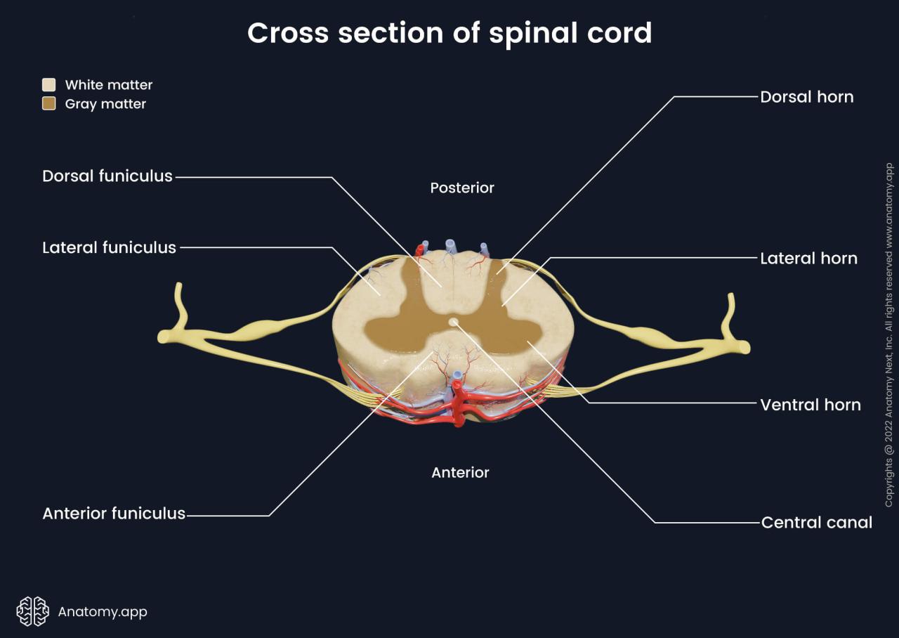
spinal cord is composed of gray matter and white matter. Gray matter forms the central core of the spinal cord and contains the cell bodies of neurons. White matter surrounds the gray matter and consists of myelinated axons, which are the long, slender extensions of neurons that transmit electrical signals.The
spinal cord is divided into four regions: cervical, thoracic, lumbar, and sacral. Each region gives rise to pairs of spinal nerves that innervate specific regions of the body.The cervical region of the spinal cord gives rise to eight pairs of spinal nerves that innervate the neck, shoulders, arms, and hands.
The thoracic region gives rise to 12 pairs of spinal nerves that innervate the chest, abdomen, and back. The lumbar region gives rise to five pairs of spinal nerves that innervate the lower back, buttocks, and legs. The sacral region gives rise to five pairs of spinal nerves that innervate the pelvis, perineum, and feet.
Functions of the Spinal Cord, Spinal cord and spinal nerves quiz
The spinal cord has a number of important functions, including:
- Motor function:The spinal cord transmits motor signals from the brain to the muscles, allowing for voluntary movement.
- Sensory function:The spinal cord transmits sensory signals from the body to the brain, allowing us to feel pain, temperature, and other sensations.
- Reflex function:The spinal cord controls a number of reflexes, which are automatic responses to stimuli.
- Autonomic function:The spinal cord helps to regulate the body’s autonomic functions, such as heart rate, blood pressure, and digestion.
Labeled Sections of the Spinal Cord
The following table shows the labeled sections of the spinal cord:
| Section | Description |
|---|---|
| Gray matter | Central core of the spinal cord containing the cell bodies of neurons. |
| White matter | Surrounds the gray matter and consists of myelinated axons. |
| Dorsal horn | Posterior portion of the gray matter that receives sensory signals. |
| Ventral horn | Anterior portion of the gray matter that sends out motor signals. |
| Dorsal root | Carries sensory signals into the spinal cord. |
| Ventral root | Carries motor signals out of the spinal cord. |
| Spinal nerve | Formed by the union of a dorsal root and a ventral root. |
Spinal Nerves: Spinal Cord And Spinal Nerves Quiz
Spinal nerves are mixed nerves that emerge from the spinal cord and transmit sensory and motor information to and from the body. They play a crucial role in communication between the central nervous system and the peripheral nervous system.
Each spinal nerve arises from the spinal cord through two roots: a dorsal (posterior) root and a ventral (anterior) root.
Dorsal and Ventral Roots
The dorsal rootcarries sensory information from the body to the spinal cord. It contains sensory neurons that receive stimuli from the skin, muscles, joints, and internal organs.
The ventral rootcarries motor information from the spinal cord to the body. It contains motor neurons that innervate muscles, allowing for voluntary movement.
The dorsal and ventral roots join together just outside the spinal cord to form a spinal nerve.
Spinal Nerves and Innervation
There are 31 pairs of spinal nerves in the human body, each corresponding to a specific level of the spinal cord.
| Spinal Nerve | Innervation Area |
|---|---|
| Cervical (C1-C8) | Neck, head, upper limbs |
| Thoracic (T1-T12) | Chest, abdomen, back |
| Lumbar (L1-L5) | Lower back, abdomen, legs |
| Sacral (S1-S5) | Pelvis, buttocks, legs |
| Coccygeal (Co1) | Tailbone area |
Spinal Reflexes
Spinal reflexes are involuntary, rapid responses to stimuli that occur at the level of the spinal cord, without involving the brain. They play a crucial role in protecting the body from harm and maintaining homeostasis.
A simple spinal reflex arc, the fundamental unit of a spinal reflex, consists of five components:
- Receptor: Detects the stimulus.
- Sensory neuron: Transmits the signal from the receptor to the spinal cord.
- Integration center: Processes the signal in the spinal cord and determines the appropriate response.
- Motor neuron: Transmits the response signal from the spinal cord to the effector.
- Effector: Responds to the signal, such as a muscle contracting or a gland secreting.
Common spinal reflexes include:
- Patellar reflex (knee-jerk reflex): Tapping the patellar tendon below the kneecap causes the knee to extend.
- Achilles reflex (ankle jerk reflex): Tapping the Achilles tendon behind the ankle causes the foot to plantarflex (point downward).
- Babinski reflex: Stroking the sole of the foot from heel to toe causes the toes to fan out (in infants, this is a normal reflex; in adults, it indicates damage to the spinal cord or brain).
Clinical Significance
Spinal cord and spinal nerve injuries can have profound clinical implications, affecting various aspects of an individual’s health and well-being.
Types of Spinal Cord Injuries
Spinal cord injuries can be classified based on the severity and location of the damage. The most common types include:
- Complete injury:All sensory and motor function is lost below the level of the injury.
- Incomplete injury:Some sensory and/or motor function is preserved below the level of the injury.
- Tetraplegia:Injury to the cervical spine, resulting in paralysis of all four limbs.
- Paraplegia:Injury to the thoracic or lumbar spine, resulting in paralysis of the lower limbs.
Diagnostic Tests
Various diagnostic tests are used to assess spinal cord and nerve function, including:
- Magnetic resonance imaging (MRI):Provides detailed images of the spinal cord and nerves.
- Computed tomography (CT) scan:Uses X-rays to create cross-sectional images of the spine.
- Electromyography (EMG):Measures electrical activity in muscles to assess nerve function.
- Nerve conduction studies:Measure the speed and amplitude of nerve impulses to assess nerve function.
Questions Often Asked
What is the function of the spinal cord?
The spinal cord serves as a vital communication pathway between the brain and the rest of the body, transmitting sensory information to the brain and motor commands from the brain to the muscles.
How many spinal nerves are there?
There are 31 pairs of spinal nerves that emerge from the spinal cord, providing sensory and motor innervation to different regions of the body.
What is a spinal reflex?
A spinal reflex is an involuntary, rapid response to a stimulus that occurs at the level of the spinal cord, without involving the brain.